



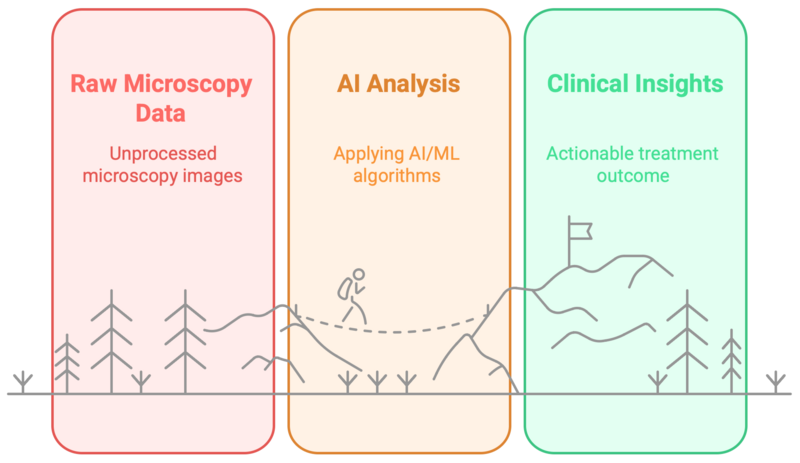
Our group is part of the Berlin Initiative for Applied Foundation Model Research (Appl-FM), a five-year programme running from 2024 to 2029. We work on a problem that sits between microbiology and machine learning: microscopy produces enormous quantities of image data, but converting that data into measurements that can support a biological claim still depends heavily on manual work.
We build pipelines that take raw fluorescence micrographs, segment every individual cell, extract quantitative morphological descriptors, and feed those descriptors into models that can classify, compare, or predict. The biology and the computation are developed together rather than handed back and forth, which is the part that usually decides whether a pipeline survives contact with real data.

What makes it difficult
Three problems recur across our datasets, and much of our methodological work targets them directly.
Class imbalance. Biologically interesting cells are often rare. A model trained naively on such data learns to predict the majority class and reports excellent accuracy while being useless.
Annotation cost. Expert-labelled microscopy data is expensive to produce. We use foundation models adapted to biomedical imaging, together with active learning strategies that choose which images are worth annotating, so that a limited expert budget goes where it changes the model most.
Validation that holds up. Cells from the same image are not independent samples. Without grouped cross-validation and held-out validation at the level of image, strain or experiment, apparent performance reflects memorisation rather than generalisation. We treat validation design as part of the experiment, not as a step at the end.
Research directions
Antimicrobial resistance. We ask whether resistance-associated phenotypes can be detected from bacterial morphology in cells grown without antibiotic exposure, which would remove the drug-exposure step from susceptibility assessment. Clinical isolates are complemented by strains in which resistance is evolved under controlled laboratory conditions, allowing comparison against a fixed genetic background.
Linking morphology to physiological state. Beyond resistance, we are interested in what else single-cell shape encodes: growth phase, stress response, and the effect of antimicrobial surfaces and biofilm inhibitors developed elsewhere in the AG Grohmann group.
Methodology. Segmentation model adaptation, descriptor design, imbalance-aware training, and uncertainty estimation for biological decision-making.
